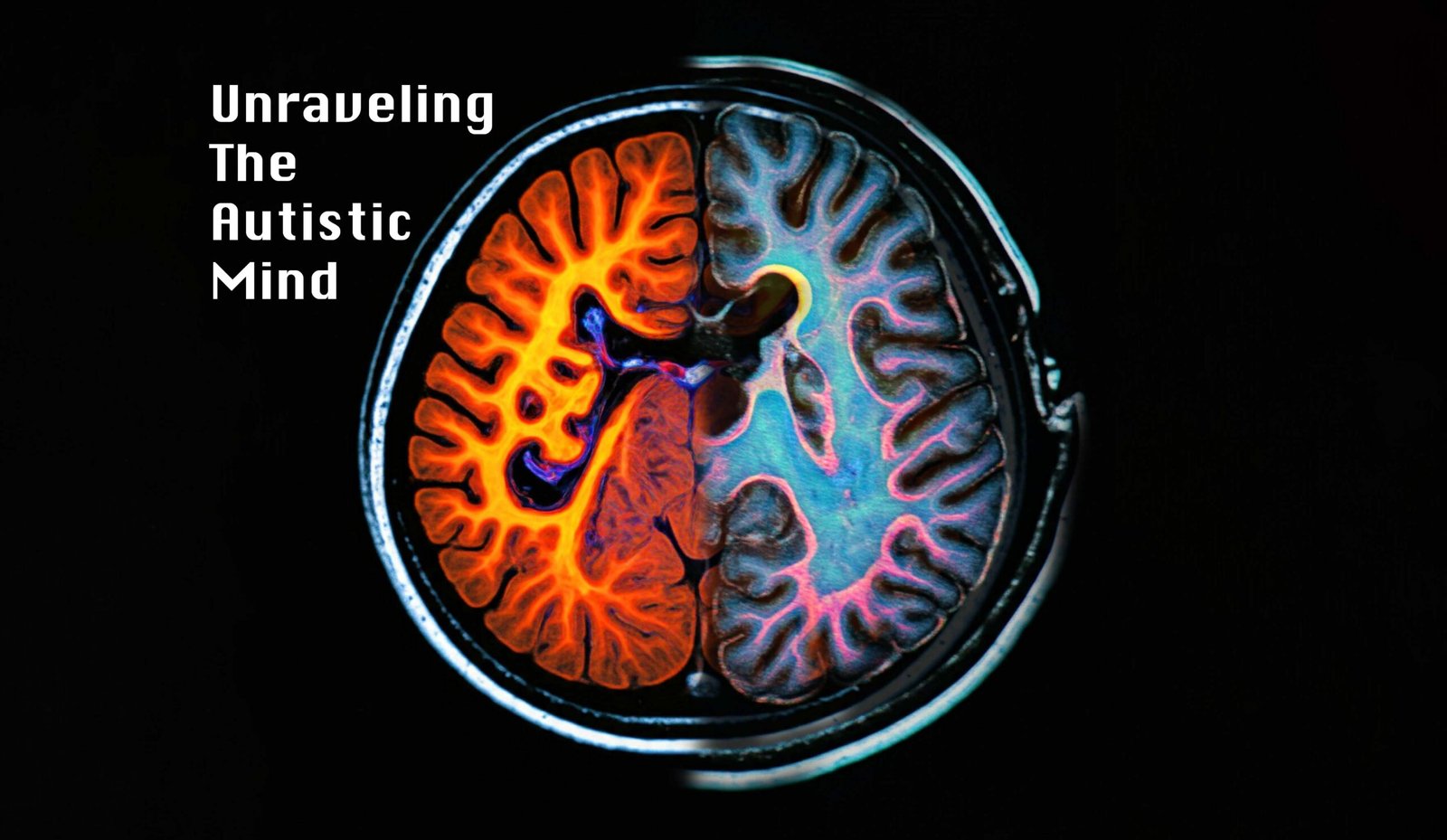
Imagine a world where colors are too bright, sounds are too loud, and every social interaction feels like navigating a complex maze. This is the reality for many individuals with Autism Spectrum Disorder (ASD). But what if we could peer into their minds, not with a single lens, but with a kaleidoscope of imaging techniques? Welcome to the fascinating world of multimodal neuroimaging in ASD research.
Autism Spectrum Disorder (ASD) is a complex neuro-developmental condition characterized by significant social impairments and restricted repetitive behaviors. The prevalence of ASD is notably higher in males than in females, with recent estimates suggesting a ratio of approximately 4:1. Understanding the neurobiological underpinnings of social deficits in ASD is crucial for developing effective interventions and support systems. Despite advancements in neuroimaging techniques, robust imaging biomarkers specific to ASD remain elusive, primarily due to the disorder’s neurobiological complexity and limitations inherent in single-modality research.Recent studies have indicated that both gray matter volume (GMV) and white matter (WM) functional activity play critical roles in social cognition and behavior. While functional MRI (fMRI) has revealed atypical brain activity in regions associated with social cognition, structural changes in both GMV and WM have also been documented. However, most research has focused on single modalities, leaving a gap in understanding how these modalities interact to reflect the complexities of social behavior in ASD.
Multimodal Fusion Analysis
To bridge this gap, a recent study employed a supervised multimodal fusion analysis to investigate the neural patterns associated with social deficits in male ASD patients. This approach integrated GMV from structural MRI and fractional amplitude of low-frequency fluctuations (WM-fALFF) from resting-state fMRI. The study utilized data from the Autism Brain Imaging Data Exchange (ABIDE) project, which included a substantial cohort of ASD and healthy control participants.The researchers aimed to explore the relationship between brain patterns and social impairments as measured by the Social Responsiveness Scale (SRS), which assesses various dimensions of social behavior. By employing a two-way supervised multimodal MRI feature fusion model, the study sought to identify neural correlates of social deficits across multiple domains, including social awareness, cognition, communication, motivation, and mannerisms.
The Autism Enigma
For decades, autism has been a puzzle that scientists, clinicians, and families have been trying to solve. It’s a condition that affects about 1 in 36 children in the United States, with a striking gender disparity – boys are four times more likely to be diagnosed than girls. But why? What’s happening in the intricate landscape of the autistic brain that creates such a unique way of perceiving and interacting with the world?
Traditionally, researchers have used individual brain imaging techniques to study ASD. It’s like trying to understand a symphony by listening to just one instrument. But the brain, in all its complexity, deserves a full orchestra of imaging methods. Enter multimodal neuroimaging – a revolutionary approach that combines multiple imaging techniques to create a more comprehensive picture of the autistic brain.
New Findings on Autistic Study
Validation Across Sites: The study employed a leave-one-site-out validation strategy, demonstrating the robustness and generalizability of the results across different cohorts. This validation reinforces the reliability of the identified neural patterns associated with social impairments in ASD.
Salience Network and Limbic System: The study found consistent involvement of the salience network (SAN) and limbic system across all social impairment domains. These regions are crucial for processing social stimuli and emotional responses. Abnormalities in these areas may contribute to the characteristic social deficits observed in individuals with ASD.
Divergent WM Functional Activity: The analysis revealed that WM-fALFF exhibited more diverse brain patterns related to different social impairments, suggesting that WM functional activity is more sensitive to the complexities of social deficits in ASD. Specific WM tracts, such as the anterior corona radiata and corpus callosum, were identified as significant contributors to social behavior.
Interconnected Brain Regions: The findings indicated potential interconnections between brain regions across modalities. For instance, the anterior corona radiata, which is part of the limbic system circuitry, was associated with impaired social responsiveness. This suggests that multimodal fusion can uncover relationships between different brain regions and their contributions to social behavior.
Gray Matter Meets White Matter
Our brains are composed of gray matter – the cell bodies of neurons where processing occurs – and white matter – the highways of the brain that connect different regions. Previous studies often focused on one or the other. But what if the key to understanding autism lies in the interplay between these two?
Recent research has done just that, combining measurements of gray matter volume (GMV) with a novel measure of white matter function called fractional amplitude of low-frequency fluctuations (WM-fALFF). It’s like looking at both the cities (gray matter) and the roads connecting them (white matter) on a complex map of the brain.
The Salience Network: The Brain’s Social Radar
One of the most intriguing findings from this multimodal approach is the consistent involvement of the salience network (SAN) in social deficits across the autism spectrum. Think of the SAN as the brain’s social radar – it helps us pick out what’s important in our environment, especially in social situations.
In individuals with autism, this network seems to be functioning differently. It’s as if their social radar is calibrated to a different frequency, making it challenging to navigate the nuances of social interactions that many of us take for granted.
White Matter: The Unsung Hero of Social Cognition
While gray matter has often taken center stage in brain research, this new approach has shone a spotlight on the critical role of white matter in social cognition. The study found that white matter functional activity was exquisitely sensitive to different aspects of social impairment in autism.
Specific white matter tracts, like the anterior corona radiata and corpus callosum, emerged as key players. These are like the major highways of the brain, connecting regions crucial for social understanding and behavior. In autism, it seems these highways might have some unexpected traffic patterns, potentially explaining some of the social challenges experienced by individuals on the spectrum.
A Symphony of Brain Regions
Perhaps the most beautiful revelation from this multimodal approach is the intricate dance between different brain regions. It’s not just about isolated areas functioning differently; it’s about how these regions communicate and work together – or sometimes fail to do so effectively.
This interconnectedness paints a picture of autism not as a disorder of any single brain region, but as a complex symphony where some instruments are playing to a slightly different tune.
From Lab to Life: The Promise of Personalized Interventions
The insights gained from this research aren’t just academically interesting – they hold real promise for improving the lives of individuals with autism. By understanding the unique neural signatures associated with different social challenges, we open the door to more personalized interventions.
Imagine a future where a child with autism could receive a brain scan that precisely identifies their unique neural patterns. This information could then be used to tailor therapies and support strategies to their specific needs, potentially leading to more effective outcomes.
The Road Ahead: A Future of Possibilities
As exciting as these findings are, they’re just the beginning. The field of multimodal neuroimaging in autism research is ripe with possibilities:
Longitudinal studies could help us understand how these brain patterns change over time, potentially leading to earlier diagnosis and intervention.
Expanding this research to include more female participants could shed light on the intriguing gender differences in autism prevalence and presentation.
Combining this imaging data with genetic information could provide a more complete picture of the biological underpinnings of autism.
A New Chapter in Understanding Autism
The journey into the autistic mind through multimodal neuroimaging is revealing a landscape more beautiful and complex than we ever imagined. It’s a story of interconnected brain networks, of highways and cities in the neural landscape, all working together to create the unique perception and interaction style that characterizes autism.
As we continue to unravel this neurological tapestry, we move closer to a world where the challenges of autism can be better understood and supported. It’s a future where the unique strengths of the autistic mind can be celebrated, and where every individual on the spectrum can find their place in the rich, diverse symphony of human neurodiversity.
The next chapter in this fascinating story is yet to be written. But with each brain scan, each study, and each breakthrough, we’re turning the page to a deeper understanding of the beautiful complexity of the human mind.
References: Wei, L., Xu, X., Su, Y., Lan, M., Wang, S., & Zhong, S. (2024). Abnormal multimodal neuroimaging patterns associated with social deficits in male autism spectrum disorder. Human Brain Mapping, 45, e70017. https://doi.org/10.1002/hbm.70017
Calhoun, V. D., & Sui, J. (2016). Multimodal fusion of brain imaging data: A key to finding the missing link(s) in complex mental illness. Biological Psychiatry: Cognitive Neuroscience and Neuroimaging, 1, 230–244. This article emphasizes the importance of multimodal imaging approaches in understanding complex mental health disorders, including autism, and supports the use of multimodal fusion in the study of ASD.


